anatomy of neck
Color X-Ray of the Cervical Spine showing Degenerative Disc Disease. 9 Images about Color X-Ray of the Cervical Spine showing Degenerative Disc Disease : Lymphatic system | 19th-century wax anatomical model showing… | Flickr, Normal flexion and extension cervical spine x-rays | Image and also Osteoid osteoma | Image | Radiopaedia.org.
Color X-Ray Of The Cervical Spine Showing Degenerative Disc Disease
 www.pinterest.com
degenerative disc disease cervical spine neck ray ddd pain mri spinal arthritis cord disk showing lumbar nerve symptoms c5 surgery
Image | Radiopaedia.org
 radiopaedia.org
metatarsal fracture 5th stress fifth radiopaedia proximal modality oblique ray version case
Image | Radiopaedia.org
 radiopaedia.org
shoulder normal ray dislocation axial rays radiopaedia axillary views orthobullets cuff rotator xr version frontal
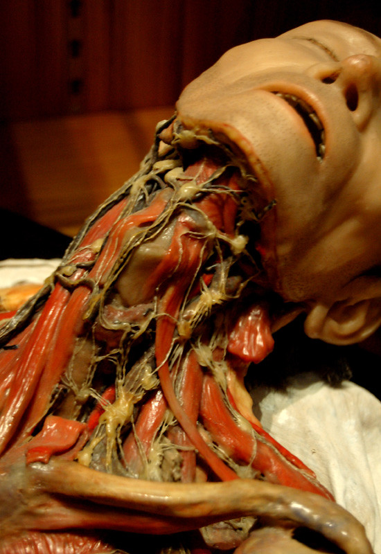
Lymphatic System | 19th-century Wax Anatomical Model Showing… | Flickr
 www.flickr.com
anatomy wax lymphatic system human lymph nodes museum body neck anatomical medical century 19th scientific sculpture animal showing sistema linfatico
Osteoid Osteoma | Image | Radiopaedia.org
 radiopaedia.org
osteoma osteoid radiopaedia ray cortical bone wikidoc case version
Medial Plantar Muscles Of The Foot: Anatomy | Kenhub
foot anatomy kenhub muscles plantar medial muscle ankle plantaris sole body diagram human sink 3d nerve study tibial anatomie read
Cranial Sutures - The Sutures Of The Skull | Kenhub
kenhub skull suture occipital anatomy lambdoid superior sutures linea nuchal line nuchalis posterior bone cranial bones joints accessory sutura sphenoid
Parapharyngeal And Retropharyngeal Spaces: Anatomy | Kenhub
carotid artery common anatomy retropharyngeal parapharyngeal space spaces pharynx dorsal kenhub skull variations anatomical
Normal Flexion And Extension Cervical Spine X-rays | Image
 radiopaedia.org
flexion radiopaedia spine
Metatarsal fracture 5th stress fifth radiopaedia proximal modality oblique ray version case. Osteoid osteoma. Shoulder normal ray dislocation axial rays radiopaedia axillary views orthobullets cuff rotator xr version frontal