anatomy of the chest wall
Hypertrophy Left Ventricle with Thickened Wall - Chest Case Studies. 9 Images about Hypertrophy Left Ventricle with Thickened Wall - Chest Case Studies : Chapter 2. Anterior Thoracic Wall | The Big Picture: Gross Anatomy, Image | Radiopaedia.org and also Pulmonary hamartoma | Image | Radiopaedia.org.
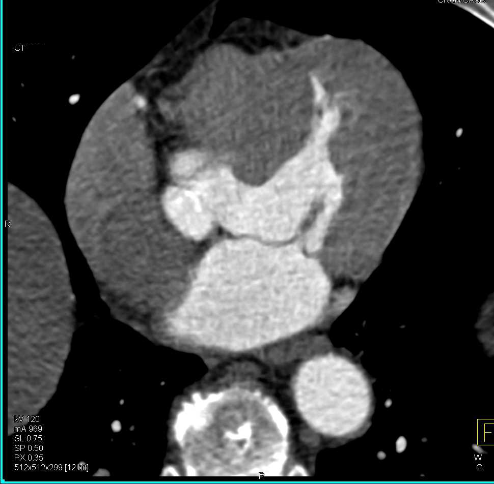
Hypertrophy Left Ventricle With Thickened Wall - Chest Case Studies
 www.ctisus.com
hypertrophy ctisus ventricle
Thoracic Cavity - Wikidoc
 www.wikidoc.org
thoracic cavity thorax lungs left sympathetic trunk among seen diaphragm joints wikidoc removed others plexus communicans ramus pulmonary breathing ventilation
Blood Vessels - Veins In Arm & Shoulder
 www.purposegames.com
blood vessels veins arm shoulder game purposegames
Septic Pulmonary Emboli And Chest Wall Abscess | Image | Radiopaedia.org
 radiopaedia.org
septic chest pulmonary emboli abscess ct radiopaedia picc line vein version
Chapter 2. Anterior Thoracic Wall | The Big Picture: Gross Anatomy
 accessmedicine.mhmedical.com
thoracic basicmedicalkey accessmedicine mhmedical
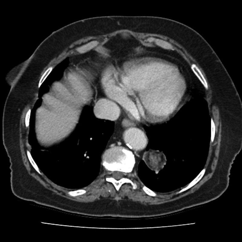
Pulmonary Hamartoma | Image | Radiopaedia.org
 radiopaedia.org
hamartoma pulmonary radiopaedia lung
Frog Abdominal And Chest Cavities
 www.purposegames.com
frog chest abdominal cavities
Shoulder Anatomy - Medical Art Library
 medicalartlibrary.com
anatomy shoulder diagram medical muscles neck bones basic muscle clavicle bone figure rotator cuff upper peeve fail medicine pet tendons
Image | Radiopaedia.org
 radiopaedia.org
radiopaedia chest anatomy silhouette contours radiology normal cardiac thorax radiography ray quiz contour pulmonary pa medical cardiovascular access figure pass
Pulmonary hamartoma. Blood vessels. Septic chest pulmonary emboli abscess ct radiopaedia picc line vein version