anatomy of tonsil
Surgical Anatomy of the Tonsils | IntechOpen. 9 Images about Surgical Anatomy of the Tonsils | IntechOpen : Waldeyer’s Ring: Definition, anatomy and pathology | Kenhub, Lateral Perspective of Brainstem and Cerebellum with Venous Anatomy and also Lateral Perspective of Brainstem and Cerebellum with Venous Anatomy.
Surgical Anatomy Of The Tonsils | IntechOpen
 www.intechopen.com
tonsils anatomy surgical intechopen figure
How To Remove Tonsil Stones At Home - Tonsil Stones Removal Home Method
 www.youtube.com
tonsil stones remove removal
Torus Tubarius – Earth's Lab
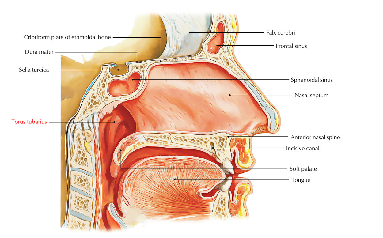 www.earthslab.com
palate soft torus incisive sella foramen cecum canal turcica tongue nasal cavity anatomy tube turcia nerve palatine eustachian thyroid sellar
Waldeyer’s Ring: Definition, Anatomy And Pathology | Kenhub
nasal ring medial palatine bone tonsil anatomy pharyngeal kenhub posterior adenoids cavity ostium tonsils tonsilla torus tubal waldeyer spine location
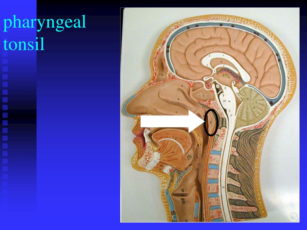
PPT - Lab Ex. 48: Lymphatic System PowerPoint Presentation, Free
 www.slideserve.com
tonsil pharyngeal lymphatic system presentation lab ex ppt powerpoint
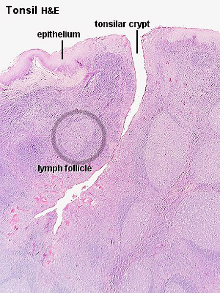
File:Tonsil Histology 01.jpg - Embryology
 embryology.med.unsw.edu.au
histology tonsil embryology
Lateral Perspective Of Brainstem And Cerebellum With Venous Anatomy
 www.neurosurgicalatlas.com
brainstem cerebellum venous neuroanatomy
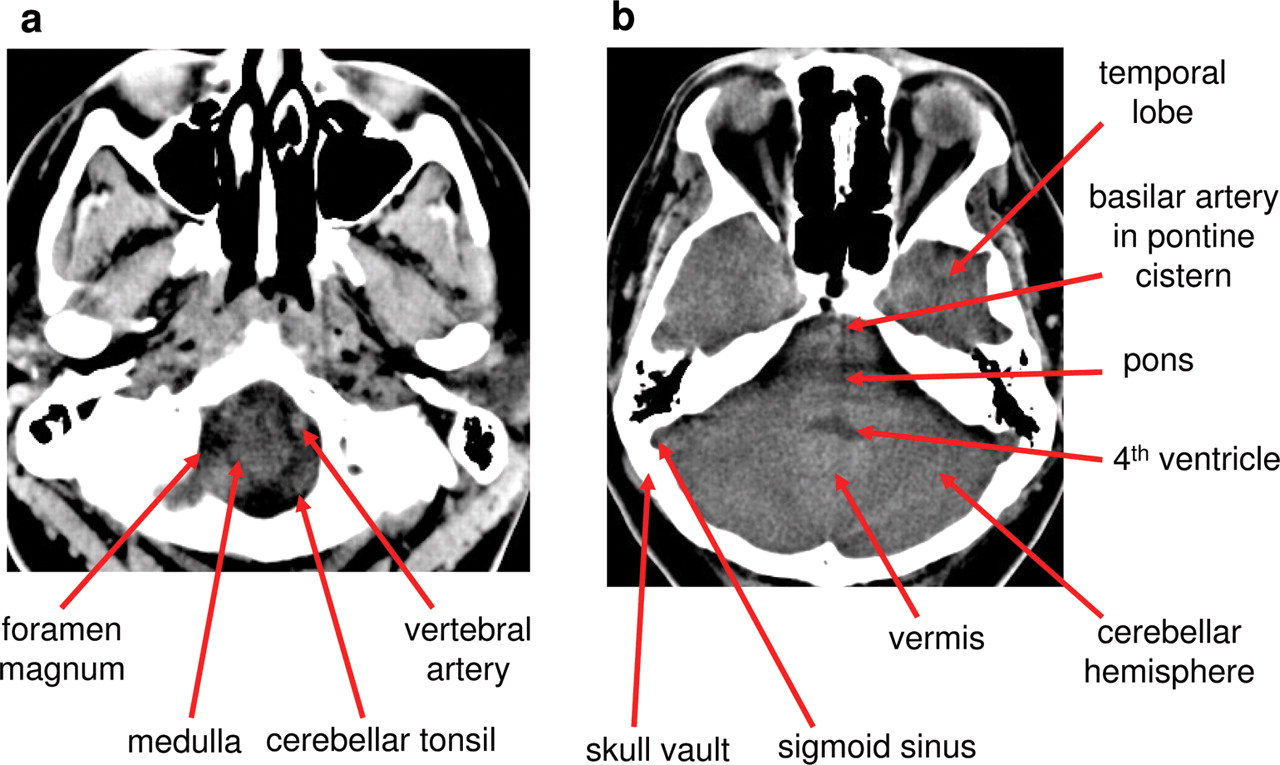
Normal Anatomy Of The Brain On CT And MRI With A Few Normal Variants
 pn.bmj.com
ct brain anatomy normal mri variants powerpoint figure tab open
Pictures Of Incision And Drainage (I&D) Of Peritonsillar Abscess
 prosites-otohouston.homestead.com
abscess peritonsillar drainage tonsil incision pus left displacing inferiorly uvula follicles surgical were right
Nasal ring medial palatine bone tonsil anatomy pharyngeal kenhub posterior adenoids cavity ostium tonsils tonsilla torus tubal waldeyer spine location. How to remove tonsil stones at home. Palate soft torus incisive sella foramen cecum canal turcica tongue nasal cavity anatomy tube turcia nerve palatine eustachian thyroid sellar