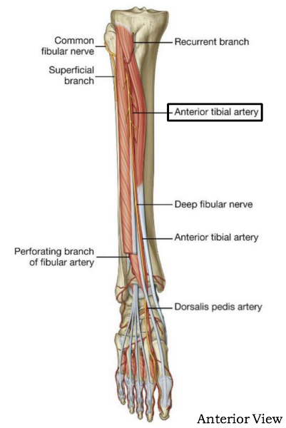
anterior tibial artery diagram
Knee (femoral–tibial) dislocation - Musculoskeletal Medicine for. 9 Images about Knee (femoral–tibial) dislocation - Musculoskeletal Medicine for : Dorsalis pedis artery – Definition, Location, Anatomy, Function, Treat foot ulcers by using the angiosome concept | Peripheral Intervention and also Anterior Drawer.
Knee (femoral–tibial) Dislocation - Musculoskeletal Medicine For
 www.orthopaedicsone.com
knee dislocation dislocations artery patellar dislocated tibial ligament surgery recovery femoral treatment symptoms types injuries injury popliteal leg reduction arterial
What Is The Splenic Artery? (with Pictures)
 www.wisegeek.com
artery splenic arteries blood organs femoral body critical abdomen internal complex major supplies system which
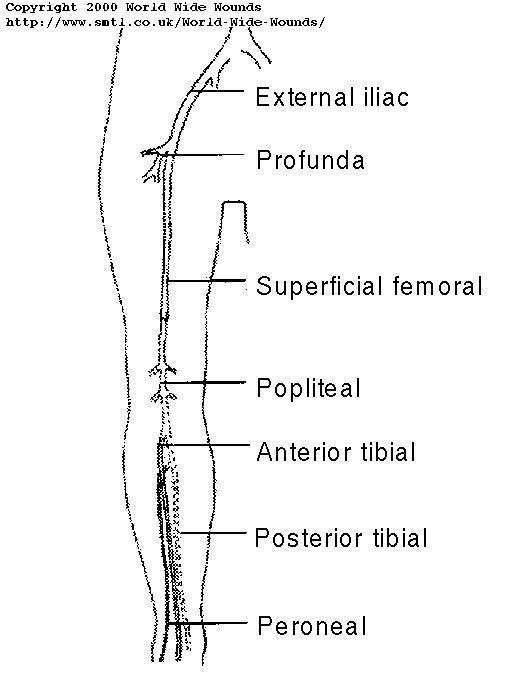
RadiologySpirit: Assessing The Lower-limb Arteries
 radiologyspirit.blogspot.com
lower arteries limb duplex extremity arterial pulse leg anatomy worksheet main doppler weak peripheral veins coloring absent figure radiologyspirit imaging
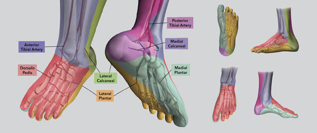
Treat Foot Ulcers By Using The Angiosome Concept | Peripheral Intervention
 www.cookmedical.eu
intervention foot peripheral anterior tibial concept ulcers artery treat using cookmedical descends
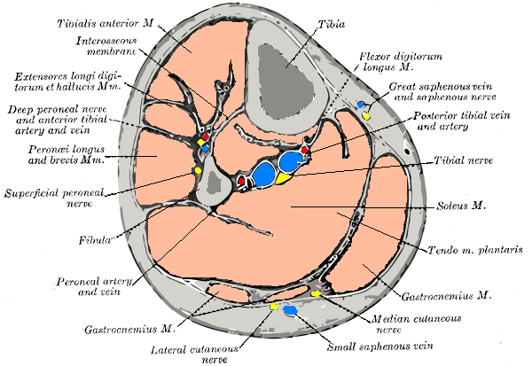
Peroneus Brevis - Wikidoc
 www.wikidoc.org
leg posterior anatomy anterior muscle nerve tibial artery saphenous vein section brevis axial superficial peroneus lower fibular gastrocnemius tibialis interosseous
Anterior Drawer
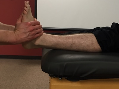 www.thestudentphysicaltherapist.com
test tunnel tarsal syndrome squeeze anterior drawer ankle talar foot special instability tests laxity syndesmotic df tilt compression
GSU Blood Vessels Of The Lower Limb Flashcards | Easy Notecards
 www.easynotecards.com
artery pedis dorsalis limb vessels gsu blood lower ankle easynotecards
Dorsalis Pedis Artery – Definition, Location, Anatomy, Function
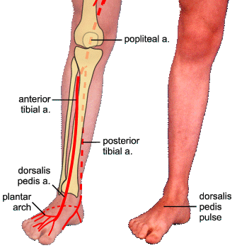 bodterms.weebly.com
dorsalis pedis artery arterial
Superficial Peroneal Nerve - Meddic
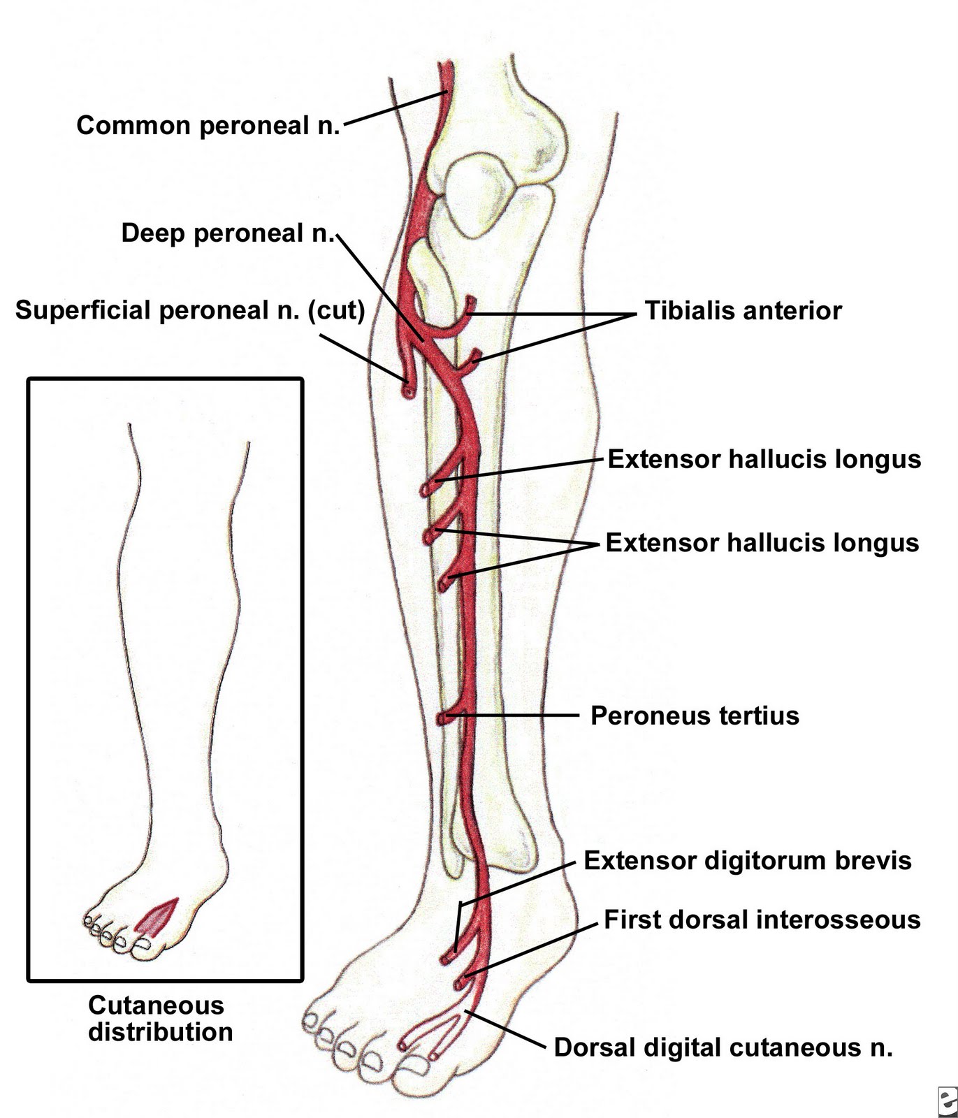 meddic.jp
peroneal peroneus profundus nervus nerves superficial fisioterapi
Knee dislocation dislocations artery patellar dislocated tibial ligament surgery recovery femoral treatment symptoms types injuries injury popliteal leg reduction arterial. Lower arteries limb duplex extremity arterial pulse leg anatomy worksheet main doppler weak peripheral veins coloring absent figure radiologyspirit imaging. Test tunnel tarsal syndrome squeeze anterior drawer ankle talar foot special instability tests laxity syndesmotic df tilt compression