axis bone anatomy
Longitudinal temporal bone fracture | Image | Radiopaedia.org. 9 Images about Longitudinal temporal bone fracture | Image | Radiopaedia.org : Atlas C1 and Axis C2 vertebrae, posterior view with labels… | Flickr, Dens (Odontoid Process) – Earth's Lab and also The Radiology Assistant : Temporal bone - Anatomy 2.0.
Longitudinal Temporal Bone Fracture | Image | Radiopaedia.org
 radiopaedia.org
fracture bone temporal longitudinal coronal radiopaedia ct mastoid contrast version non brain
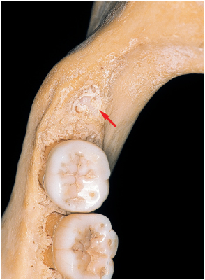
6. Surgical Protocol: Basic Principles | Pocket Dentistry
 pocketdentistry.com
retromolar molar triangle bone mandibular protocol surgical principles basic third extends cusp exposes fenestration arch dental along left pocketdentistry
Knee Ultrasound
 www.fpnotebook.com
ultrasound knee tendon quadriceps patella fpnotebook ii popliteal
Bone Anatomy And Function
 www.theodorideskneesurgeon.com
axial appendicular labeled labelled
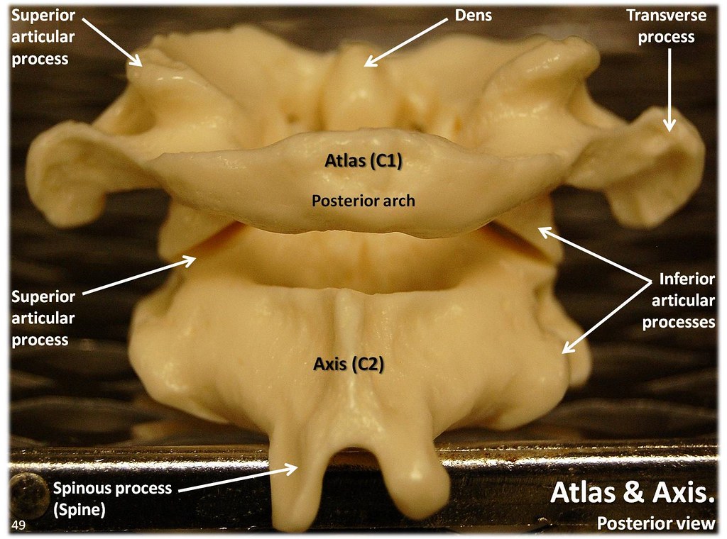
Atlas C1 And Axis C2 Vertebrae, Posterior View With Labels… | Flickr
 www.flickr.com
c1 atlas axis c2 vertebrae posterior axial skeleton labels visual flickr
Forces On Bone - Compression, Tension And Shear | Bone And Spine
 boneandspine.com
shear
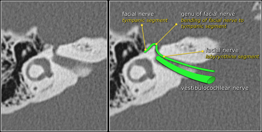
The Radiology Assistant : Temporal Bone - Anatomy 2.0
 radiologyassistant.nl
temporal bone anatomy nerve facial segment canal radiology petrous labyrinthine right coming axis angles geniculate auditory sharply nearly internal forward
Kandace's Anatomy Blog: Bones, Bones, Bones!!!
 kandaceanatomyblog.blogspot.com
fracture pediatric transverse femoral fractures femur shaft orthobullets bone bones kandace anatomy example open
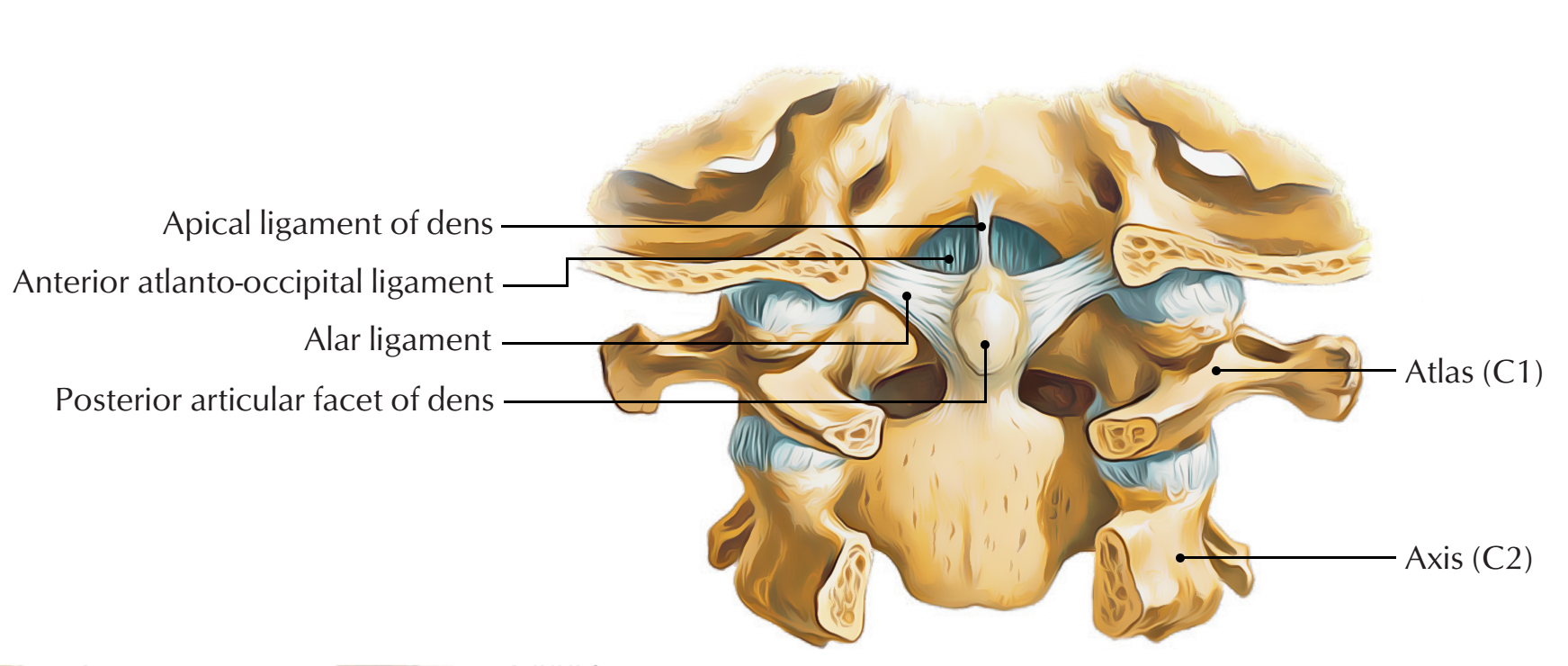
Dens (Odontoid Process) – Earth's Lab
 www.earthslab.com
dens odontoid ligaments process alar apical attached transverse magnum foramen anatomy neck craniocervical below structure upper inclined borders towards edge
Longitudinal temporal bone fracture. Bone anatomy and function. Axial appendicular labeled labelled